Title : Challenging quality assessment in medical imaging with sober newtorks. Application to MRI for stroke diagnosis.
Partners : IBISC (univ Evry, université Paris-Saclay), centre hospitalier sud-francilien (CHSF)
Basic AI and Data Science : statistical training in big dimensions
Specialized ML and AI : signal, image, vision
Application domain : precision medecine, imagery by MR
Keywords: deep learning, imagerie multi-modale, tensor decomposition, machine learning, deep tech, neuroimaging, precision medicine, stroke
total duration of internship 6 months (postgraduate)
Working period : 2024/02/01 to 2024/09/01
Context and objectives
Popular deep learning algorithms such as Chat GPT are largely based on the Transformer method which relies on an huge number of parameters. Such method require extremely expensive computing power to train, and above all, a quantity of learning data which is counted in millions or billions of examples, two requirements totally inaccessible for the majority of specialized applications such as medical applications or niche market
For several years, our laboratory has been focusing on a frugal approach to machine learning. Frugal encompass mainly the parsimonious use of learning parameters and of training samples.
Automatic learning on tensor data is classically carried out by linear tensor decomposition, for example CPD/PARAFAC or Tucker [Sid17]. Recently, tensor representations have been integrated into neural networks and have enabled significant developments in deep learning, particularly in the field of images, by reducing the number of parameters to be estimated.
To increase the identifiability and interpretability of deep neural models, constraints are added, for example non-negativity, classic in a matrix and tensor learning framework [Kol08]. In deep learning, variational autoencoders have been interpreted in a non-negative matrix factorization framework, but also as a CPD tensor factorization, and even non-negative Tucker [Mar22]. Autoencoders belong to the family of generative models. They make it possible to discover latent spaces by learning an automorphism x=f(x). Their latent space can be structured in tensor form, which provides very good performance [Pan21]. It has been shown that this allows a compromise in terms of performance and interpretability, between a simple unconstrained autoencoder and a non-negative Tucker model, for different tasks (segmentation, pattern detection). However, this preliminary work leaves significant room for progress, and the properties of this type of hybrid model are still poorly understood.
The subject we are proposing is part of a long term research to create AI tools based on MRI imaging during the acute phase of brain stroke. In particular, we are working on an innovative frugal method to make the best use of information obtained through multimodal MRI, distinguishing inter-modal corroborative information from singular uni-modal information on each of the multimodal sequences of an MRI [Bra19, Kob19].
For this project, the intern will propose and develop a deep learning algorithm to identify redundant and unique information between modalities and make the best use of it. He will be supported in this task by an experienced research team.
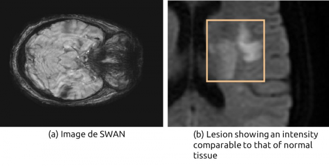
The method developed will be compared with state of the art multimodal fusion method already used in the team and apply for ischemic stroke that is caused by a blood clot (thrombus) that blocks a brain artery causing lack of oxygen brain tissue supplied by that artery (Fig. 1). MRI is the gold standard for eliminating non-vascular diagnoses because of its sensitivity and specificity in acute ischemia [Che17].
The objectives are to validate the results on a large patient database from the CHSF.
First of all, we will establish a benchmark of the different approaches. Then we will modify the constraints which structure the tensor decomposition in an auto-encoder/Tucker decomposition type model. We will evaluate and compare the characteristics of several architectures for the autoencoder. The proposed algorithms will be tested on data from several application fields currently examined in our laboratory.
Expected performance criteria [Dic45]:
Evaluating the new procedure against a referenced procedure raises many methodological difficulties. The expected performance indicators are
- the repeatability of the (deterministic) segmentation process in a degraded situation or not,
- the efficiency of the tool to be tested on a ground truth basis and quantified with DICE [3] to measure performance in segmentation,
- a speed of execution of a few minutes.
continuation of thesis
This work could continue in thesis (1) by comparing the performances of the representation in the temporal, time-frequency, time-scale domains (2) by applying these tensor decompositions on Boltzmann machines (DB networks and diffusion model) (3) by studying the influence of the network structure of the underlying phenomenon on the signal representation. Industrial collaborations are possible.
Profile and skills required
The recruited person will be in the 3rd year of engineering school or Master’s. It will be able to understand and develop adaptive learning algorithms and to process medical dataset, index it and use it in an operational system to achieve the mission described above.
Programming skills: Python or C / C ++. A practice of Pytorch would be a plus. The practice of French is not compulsory. His(her) English is fluent. The work will be carried out at the IBISC Laboratory located on the Evry campus of the UPSaclay. IBISC develops multidisciplinary, theoretical and applied research in the field of information sciences and engineering, with a strong orientation towards health applications. The selected candidate will have the chance to work in an interdisciplinary team and with a consortium of data scientists and clinicians from the CHSF. The project is multidisciplinary, at the interface of machine learning, computer science and medicine.
Scientific and material conditions
The student will be supervised by Vincent Vigneron and Sofia Vargas, from the IBISC laboratory (Univ Évry, Université Paris-Saclay). All master machine learning, signal and image processing.
Location
The internship will be carried out in part on the two sites below
- IBISC Laboratory, UFR Sciences et Technologies, Université d’Évry, Pelvoux site, 36 rue du Pelvoux 91000 ÉVRY-COURCOURONNES
- IBISC Laboratory, IBGBI site, 23, Boulevard de France 91000 ÉVRY-COURCOURONNES
Contact
Vincent Vigneron {} and Blaise Hanczar {blaise.hanczar@univ-evry.fr}
Phone: +33 1 694 775 45
References
[Kol08] Kolda, Bader, « Tensor decompositions and applications », in: SIAM review 51.3 (2009), pp. 455–500.
[Sid17] Sidiropoulos et al. « Tensor Decomposition for Signal Processing and Machine Learning » IEEE Transactions on Signal Processing, 2017.
[Pan21] Panagakis et al. « Tensor Methods in Computer Vision and Deep Learning » Proceedings of the IEEE, https://doi.org/10.1109/JPROC.2021.3074329
[Mar22] Marmoret, « Unsupervised Machine Learning Paradigms for the Representation of Music Similarity and Structure », thèse IMT Atlantique, 2022.
[Bra19] Ikram Brahim, Dominique Fourer, Vincent Vigneron, and Hichem Maaref. Deep Learning Methods for MRI Brain Tumor Segmentation: a comparative study. In 9th IEEE International Conference on Image Processing Theory, Tools and Applications (IPTA 2019), Istanbul, Turkey, November 2019.
[Che17] Liang Chen, Paul Bentley, and Daniel Rueckert. Fully automatic acute ischemic lesion segmentation in DWI using convolutional neural networks. NeuroImage: Clinical, 15:633 – 643, 2017.
[Dic45] Lee R. Dice. Measures of the amount of ecologic association between species. Ecology, 26(3):297–302, 1945.
[Kob19] J Kobold, V Vigneron, H Maaref, M Aghasaryan, C Alecu, N Chausson, Y L’hermitte, D. Smadja, and E. Läng. Stroke Thrombus Segmentation on SWAN with MultiDirectional U-Nets. In 9th IEEE International Conference on Image Processing Theory, Tools and Applications (IPTA 2019), Istanbul, Turkey, Nov 2019.
- Date de l’appel : 01/12/2023
- Statut de l’appel : Pourvu
- Contacts : Vincent VIGNERON (PR Univ. Évry, IBISC équipe SIAM), Blaise HANCZAR (PR Univ Evry; IBISC équipe AOB@S), vincentDOTvigneronATuniv-evryDOTfr, blaiseDOThanczarATuniv-evryDOTfr
- Sujet de stage niveau Master 2 (format PDF)
- Web équipe SIAM
- Web équipe AROB@S


